PDA detector optics

PDA detector optics
Topic LCI-USG-0211, Last Updated 11/07/2024

The PDA detector optics bench passes light through the solution under study and then separates it for analysis.

The detector is an ultraviolet/visible light (UV/Vis) spectrophotometer. With an array of 518 photodiodes, the detector operates within a range of 190 to 800 nm.

The figure below illustrates the light path through the optics assembly of the detector.
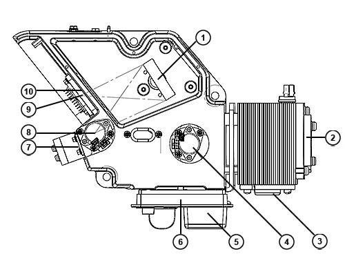
Figure 1: Optics assembly light path
Grating – Disperses light into bands of wavelengths and focuses them onto the plane of the photodiode array.
M1 mirror – Focuses light from the deuterium source lamp.
Lamp – Deuterium source lamp.
Filter flag/shutter – Flag positions for measuring open (sample) and blocked (dark) beam energies and a third for wavelength verification.
Cuvette holder - Keeps the cuvette stable and correctly aligned in the detector and allows light to pass through the sample for precise analysis.
Flow cell – Houses the segment of the flow path (containing eluent and sample) through which the polychromatic light beam passes.
Spectrograph mirror and mask – The mirror focuses light transmitted through the flow cell onto the slit at the entrance to the spectrographic portion of the optics. The mirror mask defines the size of the beam at the grating.
Variable slit - Determines the resolution and amount of light striking the PDA sensor. Smaller slit widths result in a smaller, sharper image that yields improved resolution, but with lower light throughput. Larger slit widths have higher light throughput, which can improve sensitivity and noise at the expense of lower resolution. The width of the slit is variable, but the default value is 50 µm.
Photodiode array (PDA) sensor - A linear array of 518 photodiodes, 50 µm in width, that measures the intensity of the dispersed light as a function of position on the sensor. Calibration enables the position data to be converted to wavelength to yield spectral data.
Order filter – Reduces the contribution of second-order diffraction of UV light (less than 370 nm) to the light intensity observed at visible wavelengths (greater than 345 nm).

Need Help? Connect with a Waters expert. 


To avoid risk of personal injury and to prevent damage to laboratory equipment, always operate your Waters products in accordance with the applicable operational and safety information, your organization’s standard operating procedures, and local regulations.

Refer to the English version of this page for the latest information.

Back To Top Back To Top